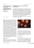
[en] The perifollicular and interfollicular areas of normal skin may look similar. However, some physiological and pathological processes may specifically involve a thin perifollicular rim. This review illustrates some of the methods available for highlighting the rim of the perifollicular epidermal unit. Non invasive methods rely on dermoscopy, ultraviolet light enhanced visualization (ULEV), skin capacitance imaging and cyanoacrylate skin surface strippings (CSSS). Conventional histology and immunohistochemistry may also show specific perifollicular features without, however, revealing the aspects highlighted by the specific non invasive methods. The clinically relevant modifications consist of pigmentary and hyperkeratotic perifollicular changes.
Disciplines :
Dermatology
Author, co-author :
Xhauflaire, Emmanuelle ; Centre Hospitalier Universitaire de Liège - CHU > Dermatopathologie
Hermanns, Jean-Francois; Centre Hospitalier Universitaire de Liège - CHU > Dermatopathologie
Pierard, Claudine ; Centre Hospitalier Universitaire de Liège - CHU > Dermatopathologie
Pierard, Gérald ; Centre Hospitalier Universitaire de Liège - CHU > Dermatopathologie
Language :
English
Title :
Highlighting the rim of the perifollicular epidermal unit.
Petit L, Piérard-Franchimont C, Saint Léger D, Loussouarn G, Piérard GE. Subclinical speckled perifollicular melanosis of the scalp. Eur J Dermatol 2002; 12: 565-8.
Deloche C, de Lacharrière O, Misciali C, Piraccini BM, Vincenzi C, Bastien P, Tardy I, Bernard BA, Tosti A. Histological features of peripilar signs associated with androgenetic alopecia. Arch Dermatol Res 2004; 295: 422-8.
Uhoda E, Piérard-Franchimont C, Petit L, Piérard GE. The conundrum of skin pores in dermocosmetology. Dermatology 2005; 210: 3-7.
Petit L, Fogouang L, Uhoda I, Smitz S, Piérard-Franchimont C. Regional variability in mottled photo-induced melanoderma in the elderly. Exp Gerontol 2003; 38: 327-31.
Piérard-Franchimont C, Al Rustom K, Ben Mosbah T, Piérard GE. Scanning electron microscopy of autoradiograms in follicular psoriasis. Emirates Med J 1989; 7: 143-7.
Satta R, Casu G, Dore F, Longinotti M, Cottoni F. Follicular spicules and multiple ulcers: cutaneous manifestations of multiple myeloma. J Am Acad Dermatol 2003; 49: 736-40.
Franchimont C, Arrese Estrada J, Ben Mosbah T, Piérard GE. Introduction à la physiologie cutanée. In: Piérard GE, Caumes E, Franchimont C, Arrese Estrada J, eds. Dermatologie tropicale. Publ. Ed Université de Bruxelles/AUPELF, 1993: 59-72.
Uhoda E, Piérard-Franchimont C, Petit L, Piérard GE. Skin weathering and ashiness in black Africans. Eur J Dermatol 2003; 13: 574-8.
Uhoda E, Petit L, Piérard-Franchimont C, Piérard GE. Ultraviolet light-enhanced visualization of cutaneous signs of carotene and vitamine A dietary deficiency. Acta Clin Belg 2004; 59: 97-101.
Hermanns JF, Petit L, Piérard-Franchimont C, Cauwenbergh G, Piérard GE. Unraveling the patterns of subclinical phaeomelanin-enriched facial hyperpigmentation. Effect of depigmenting agents. Dermatology 2000; 201: 118-22.
Lévêque JL, Querleux B. SkinChip®, a new tool for investigating the skin surface in vivo. Skin Res Technol 2003; 9: 343-7.
Piérard GE, Lévêque JL. What is SkinChip®? From silicon image sensor technology to SkinChip®. Dermatology 2004; 208: 291-2.
Uhoda E, Lévêque JL, Piérard GE. Silicon image sensor technology for in vivo detection of surfactant induced corneocyte swelling and drying. Dermatology 2005; 250: 184-8.
Xhauflaire-Uhoda E, Piérard GE. Skin capacitance imaging of acne lesions. Skin Res Technol (in press).
Marks R, Dawber RPR. Skin surface biopsy: an improved technique for the examination of the horny layer. Br J Dermatol 1971; 84: 117-23.
Piérard GE, Piérard-Franchimont C, Goffin V. Digital image analysis of microcomedones. Dermatology 1995; 190: 99-103.
Letawe C, Boone M, Piérard GE. Digital image analysis of the effect of topically applied linoleic acid on acne microcomedones. Clin Exp Dermatol 1998; 23: 56-8.
Uhoda E, Piérard-Franchimont C, Piérard GE. Comedolysis by a lipohydroxyacid formulation in acne prone subjects. Eur J Dermatol 2003; 13: 65-8.
Quatresooz P, Martalo O, Piérard GE. Differential expression of α1 (IV) and α5 (IV) collagen chains in basal cell carcinoma. J Cutan Pathol 2003; 30: 548-52.
Quatresooz P, Piérard GE. Distinct expression patterns of α1 (IV) and α5 (IV) collagen chains in cylindroma and malignant cylindroma. Int J Mol 2005; 15: 27-31.