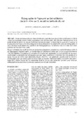
[en] Thirty equine isolated limbs have been examined by ultrasonography (7.5 MHz probes) using the frog as acoustic window. The aims of the study were to establish the ultrasonographic anatomy of the distal portion of the deep digital flexor tendon, to determine the capability to visualize podotrochlear bursa and flexor surface fibrocartilage by ultrasound and to evaluate the sensitivity of ultrasonography for the assessment of flexor surface abnormalities. Because of the relative orientation of the ultrasound beam and the deep digital flexor tendon fibers, this tendon always appears hypoechogenic on transcuneal ultrasonographic images. The flexor surface fibrocartilage is difficult to see if the podotrochlear bursa is not injected. The thickness of the normal bursa is negligible at the palmar aspect of the distal sesamoid bone. Sensitivity of ultrasonography for the detection of flexor surface irregularities was high (90,91%), sensitivity was 84,21%. Ultrasonography using the transcuneal approach could be an useful adjunct to routine radiographic examination of the foot in the assessment of flexor surface irregularities. [fr] Trente membres isolés de cheval ont été échographiés avec des sondes 7,5 MHz utilisant la fourchette comme fenêtre acoustique (voie transcunéale), afin d’étudier l’échoanatomie de la partie distale du tendon du muscle fléchisseur profond du doigt, de déterminer la capacité à visualiser la bourse podotrochléaire et le fibrocartilage de la facies flexoria de l’os sésamoïde distal par échographie et donc d’évaluer la sensibilité et la spécificité de l’échographie pour la détection des anomalies de la face palmaire de l’os sésamoïde distal.
De par l’orientation de ses fibres vis-à-vis du faisceau ultrasonore, le tendon du muscle fléchisseur profond du doigt apparaît hypoéchogène. Le fibrocartilage est difficilement visible quand la bourse podotrochléaire n’est pas injectée et l’épaisseur de la bourse non injectée est négligeable à la face palmaire de l’os sésamoïde distal. La sensibilité de l’échographie par voie transcunéale pour la détection des irrégularités de la facies flexoria de l’OSD s’est démontrée élevée (90,91%) ; sa spécificité calculée était de 84,21%. L’échographie par voie transcunéale se révèle donc un examen intéressant dans la recherche des pathologies de la facies flexoria de l’os sésamoïde distal et pourrait être combinée utilement avec l’examen radiographique du pied chez les chevaux suspects de maladie naviculaire.
Disciplines :
Veterinary medicine & animal health
Author, co-author :
Busoni, Valeria ; Université de Liège - ULiège > Département clinique des animaux de compagnie et des équidés > Imagerie médicale
Méan, Marie-Noël
Brignone, Luca
Snaps, Frédéric ; Université de Liège - ULiège > Département clinique des animaux de compagnie et des équidés > Imagerie médicale
Language :
French
Title :
Echographie de l’appareil podotrochléaire : étude in vitro sur 30 membres isolés de cheval
Alternative titles :
[en] Ultrasonography of the podotrochlear apparatus: an in vitro study on 30 equine isolated limbs
Ackerman N., Johnson J.H., Dorn C.R. Navicular disease in the horse: risk factors, radiographic changes and response to therapy. J. Am. Vet. Med. Ass. 1977, 170,183-187.
Adams O.R. Lameness in horses. Lea & Febiger: Philadelphia, 1974, 905 p.
Busoni V., Denoix J.M. Ultrasonographic examination of the podotrochlear apparatus in the horse using a transcuneal approach. Proceedings of the European Association of Veterinary Diagnostic Imaging 5th Annual Conference, 1998, O39.
Busoni V., Denoix J.M. Ultrasonography of the podotrochlear apparatus using a transcuneal approach: technique and reference images Vet. Radiol. Ultrasound, 2001, 42, 534-540.
Campbell J.R., Lee R. Radiological techniques in the diagnosis of navicular disease. Equine Vet. J., 1972, 4,135-138.
Colles C.M. Navicular disease and its treatment. In Practice, 1982, 4, 29-34.
Denoix J.M. Ultrasonographic examination in the diagnosis of joint disease. In: McIlwraith C.W., Trotter W.B. (Eds.), Joint Disease in the Horse. WB Saunders: Philadelphia, 1996, 165-202.
Denoix J.M. Joints and miscellaneous tendons. In: Rantanen N.W., McKinnon A.O. (Eds.), Equine Diagnostic Ultrasonography. Williams and Wilkins : Baltimore, 1998a, 475-514.
Denoix J.M. Symptomatologie et diagnostic du syndrome naviculaire. Proceedings de la journée de la Société des Vétérinaires Suisses, 1998b, 76-78.
Denoix J.M. The equine distal limb. An atlas of clinical anatomy and comparative imaging. Manson Publishing: London, 2000, 390 p.
Denoix J.M., Busoni V. Ultrasonography of joints and synovia. In: White N.A., Moore J.N. (Eds.), Current techniques in equine surgery and lameness. WB Saunders: Philadelphia, 1998, 643-654.
Gabriel A. Étude morphologique du petit sésamoïde du cheval. Relations éventuelles avec la pathologie. (PhD Thesis). Faculté de Médecine Vétérinaire, Université de Liège, 1997, 317p.
Hauser M.L., Rantanen N.W., Modransky P.D. Ultrasound examination of distal interphalangeal joint, navicular bursa, navicular bone and deep digital flexor tendon. Equine Vet. Sci., 1982, 2, 95-97.
Hickman J. Navicular disease - What are we talking about? Equine Vet. J., 1989, 21, 395-398.
Mackinnon A. A spreadsheet for the calculation of comprehensive statistics for the assessment of diagnostic tests and inter-rater agreement. Comput. Biol. Med., 2000, 30, 127-134.
Pool R.R., Meagher D.M., Stover S.M. Pathophysiology of navicular syndrome. Vet. Clin. Nort. Am.: Equine Pract., 1989, 5, 109-129.
Poulos P.W. Correlation of radiographic signs and histologic changes in navicular disease. Proceedings of the American Association of Equine Practitioners, 1983, 29, 241-255.
Poulos P.W., Brown A. On navicular disease in the horse. A roentgenological and Patho-anatomic Study. Part 1. Evaluation of the Flexor Central Eminence. Vet. Radiol., 1989, 30, 50-53.
Seyrek-Intas D., Tellhelm B., Reckels F.J. Interpretation and diagnostic value of some radiological findings in the navicular bone. Proceedings of the International Veterinary Radiology Association 11th Meeting, 1997, 5.
Schechter M. Evaluation of diagnosis process. In: Troidl H. Principles and practice of research: strategies for surgical investigators. Springer-Verlag: New-York, 1986, 193-206.
Turner T.A. Diagnosis and treatment of the navicular syndrome in horses. Vet. Clin. North Am.: Equine Pract., 1989, 5, 131-144.
Verschooten F. The importance of lateromedial view for the radiographic diagnosis of navicular disease. Vet. Annual, 1990, 30, 172-180.
Wright I.M. A study of 118 cases of navicular disease: clinical features. Equine Vet. J., 1993, 25, 488-492.
Wright I.M., Kidd L., Thorp B.H. Gross, histological and histomorphometric features of the navicular bone and related structures in the horse. Equine Vet. J., 1998, 30, 220-234.