Abstract :
[en] Introduction
Dermatophytoses are superficial cutaneous mycoses observed in both humans and animals caused by filamentous fungi called dermatophytes. Human dermatophytosis is widespread and most often caused by species belonging to the genus Trichophyton. The increasing emergence of strains resistant to currently available antifungals requires the development of new treatments. In this context, a better knowledge of the pathogenesis of dermatophytosis, including the identification of fungal virulence factors such as subtilisins, is a key step for the identification of new therapeutic targets. A method for standardizing the production of dermatophyte spores to perform experimental infections has been published by our research team (Faway et al., 2021). If this method based on the use of suspensions enriched in unicellular spores allows the development of standardized infections on human epidermis reconstructed in vitro, it is not optimal to produce relevant in vivo infections. The objective of this study is to develop a new relevant mouse model of infection with a reference strain of Trichophyton benhamiae and derived deleted strains, using a new standardized inoculum and to evaluate the expression of genes coding for subtilisins during the infection process.
Methods & Materials
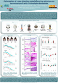
Using tBLASTn and bioinformatics analysis, 12 genes encoding subtilisins were identified in the genome of T. benhamiae. To assess the involvement of these subtilisins in the infection, the T. benhamiae IHEM 20161 strain deleted at the Ku70 locus was used to produce six strains invalidated for one or more subtilisins (∆Sub6; ∆Sub7; ∆Sub8; ∆Sub10; ∆Sub6.∆Sub10 and ∆Sub6.∆Sub8.∆Sub10). These strains were tested in a new epicutaneous mouse infection model, using a mixture of spores (1.108 CFU), germ tubes and mycelium (100 mg) as an inoculum. Kinetic monitoring of the infection was carried out by the establishment of a global clinical score based on the intensity (0 to 4) of three clinical signs, i.e. erythema, scaling and crusting, while the expression of the 12 subtilisins was evaluated by real-time quantitative PCR two and five days after infection.
Results
The inoculum used in this new in vivo model generates clearly visible skin symptoms mimicking a self-resolving natural infection, lasting more than twice as long (16 days vs 7 days) as those obtained in the past with only spores as inoculum. Clinical scores did not differ significantly between wild type and strains knocked out for one or more subtilisins. Sub1, Sub3, Sub6 and Sub10 were overexpressed by the wild type strain during the infection process, while several compensation phenomena, notably the overexpression of Sub5 by strain ∆Sub8, were observed.
Conclusion
Using a new optimized reproducible mouse model of T. benhamiae dermatophytosis allowing the study of fungal virulence factors, we show that the invalidation of subtilisin genes would induce compensatory mechanisms of overexpression of other subtilisins having a similar activity, testifying to the importance of these proteases during the infection in mice. Other ongoing research will include histological analysis, quantification of fungal genomic DNA, and analysis of the expression of other virulence factors in skin lesions.