[en] Sepsis has a profound deleterious effect on kidney functions through complex mechanisms, which involve the immune response, inflammatory pathways, intracellular dysfunction and hemodynamic instability. Those factors are difficult to discriminate in vivo. To get a better understanding of renal respiratory dysfunction, we developed an in vitro model of sepsis-induced acute kidney injury using proximal tubular epithelial cell lines (HK-2) exposed to a bacterial endotoxin (lipopolysaccharide, LPS). Using this model, our first work has demonstrated that the basal respiration of renal HK-2 cells subjected to endotoxins was altered and presented a strong decrease in the oxygen consumption rates.
Our working hypothesis of the pathophysiology of sepsis-induced AKI is based on a change in mitochondrial function that has been termed cytopathic hypoxia. A consequence of mitochondrial function alterations is an inability of the cell to use molecular oxygen for ATP production. The oxidative phosphorylation within mitochondria is interrupted because of the inhibition of cytochrome oxidase.
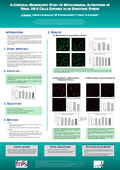
The present investigation was carried out to establish whether mitochondrial alterations might be a mechanism of renal tubular epithelial injury during sepsis. To reach this goal the mitochondrial alterations of renal HK-2 cells exposed to an endotoxic stress was studied by confocal laser-scanning microscope. Confocal microscope allowed observation of the evoked phenomena at the single cell level and in real time. More particulary, mitochondrial morphology, mitochondrial membrane potential (ΔΨm) and generation of reactive oxygen species were recorded using specific vital fluorescent probes and quantified by image processing and analysis.
Mitochondrial membrane potential is generated by the mitochondrial electron transport chain. This gradient is critical for the formation of ATP, and a fall in membrane potential is an indicator of mitochondrial dysfunction. ΔΨm was measured using the lipophilic cationic probe TMRE and it was shown that LPS produced a decrease in ΔΨm. In parallel, superoxide generation was measured by using MitoSOX which is selectively targeted to the mitochondria. There was a significant increase in mitochondrial superoxide-specific oxidation of MitoSOX when HK-2 cells were submitted to LPS.
Overall, the model of HK-2 cells exposed to LPS displays some key features of sepsis-induced acute kidney injury. The confocal microscopy study has suggested a mechanism of toxicity dependent on mitochondrial oxidant generation and mitochondrial dysfunction. Indeed, the exposure to LPS has resulted in an increased generation of superoxide and a loss of mitochondrial function probably initiated by a fall in mitochondrial potential.