Polymeric foam; Nanocomposite; X-ray microtomography; Pore characterisation; 3D image analysis
Abstract :
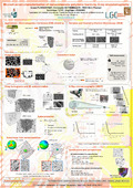
[en] Recent advances in microstructured materials have given rise to many new types of composites that exhibit original and interesting physical properties. For example, a nanocomposite made of carbon nanotubes inside a polymer matrix shows exceptional electromagnetic interference shielding effectiveness when foamed. However, the effective properties of such materials strongly depend on the shape and topology of the microstructural cells. An accurate method for investigating the cellular microstructure is X-ray microtomography (XRμT), for it is non-destructive, and it provides 3D geometric information. Although it cannot be used to observe nanofiller dispersion, it has a strong potential for cell structure characterisation.
In order to reduce the need for trial and error in tailoring these materials, our objective is to quantify, using XRμT, cellular microstructure, for two different types of foaming procedures, namely supercritical CO2 batch foaming and freeze drying, to be able to establish a link between the structure and its shielding effectiveness. The main difficulty stems from the type of material being studied: it is light, therefore hardly absorbs X-rays, cell size is small compared to the resolution capacity of the tomograph, and cell wall thickness is extremely thin in some cases, making them very hard to discern in the images. For these reasons, common image analysis tools for identifying and delimiting objects in an image prove impractical. We propose an original method that uses the 3D autocorrelation function of the tomograms to determine statistical information from these images, such as average cell size and anisotropy, without the need to binarise and segment the images.
Research Center/Unit :
Laboratory of Chemical Engineering (LGC), Faculty of Applied Sciences, ULiège ; Centre for Education and Research on Macromolecules (CERM), Faculty of Sciences, ULiège
Disciplines :
Materials science & engineering
Author, co-author :
Plougonven, Erwan ; Université de Liège - ULiège > Département de chimie appliquée > Génie chimique - Procédés et développement durable
Detrembleur, Christophe ; Université de Liège - ULiège > Centre d'études et de rech. sur les macromolécules (CERM)
Tran, Minh Phuong ; Université de Liège - ULiège > Centre d'études et de rech. sur les macromolécules (CERM)
Toye, Dominique ; Université de Liège - ULiège > Département de chimie appliquée > Génie de la réaction et des réacteurs chimiques
Léonard, Angélique ; Université de Liège - ULiège > Département de chimie appliquée > Génie chimique - Procédés et développement durable
Language :
English
Title :
Microstructure characterisation of nanocomposite polymeric foams by X-ray microtomography
Publication date :
26 March 2012
Number of pages :
A0
Event name :
6th International Symposium on Process Tomography
Event date :
from 26-03-2012 to 28-03-2012
Audience :
International
Name of the research project :
ARC project 09/14-02 - From Imaging to geometrical modelling of complex micro structured materials: Bridging computational engineering and material science