7 Tesla magnetic resonance spectroscopy estimates of GABA concentration relate to physiological measures of tonic inhibition in the human motor cortex. - 2025
7 Tesla magnetic resonance spectroscopy estimates of GABA concentration relate to physiological measures of tonic inhibition in the human motor cortex.
7 Tesla magnetic resonance spectroscopy estimates ofGABA concentration relate to physiological measures oftonic inhibition in the human motor cortex.pdf
GABA; TMS‐EEG; inhibition; magnetic resonance spectroscopy (MRS); motor cortex; neural mass model
Abstract :
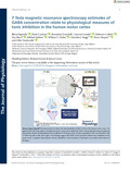
[en] GABAergic neurotransmission within the cortex plays a key role in learning and is altered in several brain diseases. Quantification of bulk GABA in the human brain is typically obtained by magnetic resonance spectroscopy (MRS). However, the interpretation of MRS-GABA is still debated. A recent mathematical simulation contends that MRS detects extrasynaptic GABA, mediating tonic inhibition. Nevertheless, no empirical data have yet confirmed this hypothesis. Here we collected ultra-high-field 7 Tesla MRS and transcranial magnetic stimulation coupled with high-density electroencephalography (TMS-hdEEG) from the motor cortex of 20 healthy participants (age 23.95 ± 6.4 years), while they were at rest. We first applied a neural mass model (NMM) to TMS-evoked potentials to disentangle the contribution of different GABAergic pools. We then assessed to which of these different pools MRS-GABA was related to by means of parametric empirical Bayesian (PEB) analysis. We found that MRS-GABA was mostly positively related to the NMM-derived measures of tonic inhibition and overall functionality of the GABAergic synapse. This relationship was reliable enough to predict MRS-GABA from NMM-GABA. These findings clarify the mesoscopic underpinnings of GABA levels measured by MRS. Our work will help fulfil the promises of MRS-GABA, enhancing our understanding of human behaviour, brain physiology and pathophysiology. KEY POINTS: GABA neurotransmission is essential for synaptic plasticity and learning (especially motor learning) and is altered in several brain disorders, such as epilepsy and stroke. Quantification of GABA in the human brain is typically obtained by magnetic resonance spectroscopy (MRS). However, the interpretation of MRS-GABA is still debated. By using a biophysical neural mass model, here we show that MRS-GABA relates to physiological measures of tonic inhibition in the human cortex.
Zanichelli, Benedetta; GIGA-Research, CRC-Human Imaging Unit, 8 allée du Six Août, Batiment B30, University of Liège, Liège, Belgium
Lamalle, Laurent ; Université de Liège - ULiège > Département de physique
Collette, Fabienne ; Université de Liège - ULiège > Département de Psychologie
Sherif, Siya ; Université de Liège - ULiège > Département d'électricité, électronique et informatique (Institut Montefiore)
Zubkov, Mikhail ; Université de Liège - ULiège > Département des sciences biomédicales et précliniques
Clarke, William T ; Wellcome Centre for Integrative Neuroimaging, FMRIB, Nuffield Department of Clinical Neurosciences, University of Oxford, Oxford, UK
Stagg, Charlotte J ; Wellcome Centre for Integrative Neuroimaging, FMRIB, Nuffield Department of Clinical Neurosciences, University of Oxford, Oxford, UK ; Medical Research Council Brain Network Dynamics Unit, University of Oxford, Oxford, UK
Maquet, Pierre ; Université de Liège - ULiège > Département des sciences cliniques > Neurologie
Vandewalle, Gilles ; Université de Liège - ULiège > Département des sciences biomédicales et précliniques
Language :
English
Title :
7 Tesla magnetic resonance spectroscopy estimates of GABA concentration relate to physiological measures of tonic inhibition in the human motor cortex.
Adams, N. E., Hughes, L. E., Rouse, M. A., Phillips, H. N., Shaw, A. D., Murley, A. G., Cope, T. E., Bevan-Jones, W. R., Passamonti, L., Street, D., Holland, N., Nesbitt, D., Friston, K., & Rowe, J. B. (2021a). GABAergic cortical network physiology in frontotemporal lobar degeneration. Brain, 144(7), 2135–2145.
Adams, N. E., Hughes, L. E., Rouse, M. A., Phillips, H. N., Shaw, A. D., Murley, A. G., Cope, T. E., Bevan-Jones, W. R., Passamonti, L., Street, D., Holland, N., Nesbitt, D., Friston, K., & Rowe, J. B. (2021b). GABAergic cortical network physiology in frontotemporal lobar degeneration. Brain, 144(7), 2135–2145.
Bachtiar, V., Near, J., Johansen-Berg, H., & Stagg, C. J. (2015). Modulation of GABA and resting state functional connectivity by transcranial direct current stimulation. eLife, 4, 1–9.
Belelli, D., Harrison, N. L., Maguire, J., Macdonald, R. L., Walker, M. C., & Cope, D. W. (2009). Extrasynaptic GABAA receptors: Form, pharmacology, and function. Journal of Neuroscience, 29(41), 12757–12763.
Bhat, R., Axtell, R., Mitra, A., Miranda, M., Lock, C., Tsien, R. W., & Steinman, L. (2010). Inhibitory role for GABA in autoimmune inflammation. Proceedings of the National Academy of Sciences, 107(6), 2580–2585.
Blicher, J. U., Near, J., Næss-Schmidt, E., Stagg, C. J., Johansen-Berg, H., Nielsen, J. F., Østergaard, L., & Ho, Y.-C. L. (2015). GABA levels are decreased after stroke and GABA changes during rehabilitation correlate with motor improvement. Neurorehabilitation and Neural Repair, 29(3), 278–286.
Bouilleret, V., Loup, F., Kiener, T., Marescaux, C., & Fritschy, J. (2000). Early loss of interneurons and delayed subunit-specific changes in GABAA-receptor expression in a mouse model of mesial temporal lobe epilepsy. Hippocampus, 10(3), 305–324.
Cao, G., Edden, R. A. E., Gao, F., Li, H., Gong, T., Chen, W., Liu, X., Wang, G., & Zhao, B. (2018). Reduced GABA levels correlate with cognitive impairment in patients with relapsing-remitting multiple sclerosis. European Radiology, 28(3), 1140–1148.
Chandra, D., Halonen, L. M., Linden, A.-M., Procaccini, C., Hellsten, K., Homanics, G. E., & Korpi, E. R. (2010). Prototypic GABAA receptor agonist muscimol acts preferentially through forebrain high-affinity binding sites. Neuropsychopharmacology, 35(4), 999–1007.
Chang, L., Jiang, C. S., & Ernst, T. (2009). Effects of age and sex on brain glutamate and other metabolites. Magnetic Resonance Imaging, 27(1), 142–145.
Chellappa, S. L., Gaggioni, G., Ly, J. Q. M., Papachilleos, S., Borsu, C., Brzozowski, A., Rosanova, M., Sarasso, S., Luxen, A., Middleton, B., Archer, S. N., Dijk, D. J., Massimini, M., Maquet, P., Phillips, C., Moran, R. J., & Vandewalle, G. (2016). Circadian dynamics in measures of cortical excitation and inhibition balance. Scientific Reports, 6, 1–13.
Chowdhury, F. A., O'Gorman, R. L., Nashef, L., Elwes, R. D., Edden, R. A., Murdoch, J. B., Barker, G. J., & Richardson, M. P. (2015). Investigation of glutamine and GABA levels in patients with idiopathic generalized epilepsy using MEGAPRESS. Journal of Magnetic Resonance Imaging, 41(3), 694–699.
Cirillo, J., Mooney, R. A., Ackerley, S. J., Barber P. A., Borges, V. M., Clarkson, A. N., Mangold, C., Ren, A., Smith, M. C., Stinear, C. M., & Byblow, W. D. (2020). Neurochemical balance and inhibition at the subacute stage after stroke. Journal of Neurophysiology, 123(5), 1775–1790.
Clarke, W. T., Bell, T. K., Emir, U. E., Mikkelsen, M., Oeltzschner, G., Shamaei, A., Soher, B. J., & Wilson, M. (2022). NIfTI-MRS: A standard data format for magnetic resonance spectroscopy. Magnetic Resonance in Medicine, 88(6), 2358–2370.
Clarke, W. T., Stagg, C. J., & Jbabdi, S. (2021). FSL-MRS: An end-to-end spectroscopy analysis package. Magnetic Resonance in Medicine, 85(6), 2950–2964.
Cuypers, K., Hehl, M., van Aalst, J., Chalavi, S., Mikkelsen, M., Van Laere, K., Dupont, P., Mantini, D., & Swinnen, S. P. (2021). Age-related GABAergic differences in the primary sensorimotor cortex: A multimodal approach combining PET, MRS and TMS. Neuroimage, 226, 117536.
Di Lazzaro, V., Capone, F., Paolucci, M., Assenza, F., Brunelli, N., Ricci, L., & Florio, L. (2016). Canonical cortical circuits: Current evidence and theoretical implications. Neuroscience and Neuroeconomics, 2016, 1.
di Lazzaro, V., & Ziemann, U. (2013). The contribution of transcranial magnetic stimulation in the functional evaluation of microcircuits in human motor cortex. Frontiers in Neural Circuits, 7, 18.
Dyke, K., Pépés, S. E., Chen, C., Kim, S., Sigurdsson, H. P., Draper, A., Husain, M., Nachev, P., Gowland, P. A., & Morris, P. G. (2017). Comparing GABA-dependent physiological measures of inhibition with proton magnetic resonance spectroscopy measurement of GABA using ultra-high-field MRI. Neuroimage, 152, 360–370.
Epperson, C. N., Haga, K., Mason, G. F., Sellers, E., Gueorguieva, R., Zhang, W., Weiss, E., Rothman, D. L., & Krystal, J. H. (2002). Cortical γ-aminobutyric acid levels across the menstrual cycle in healthy women and those with premenstrual dysphoric disorder: A proton magnetic resonance spectroscopy study. Archives of General Psychiatry, 59(9), 851–858.
Erdfelder, E., FAul, F., Buchner, A., & Lang, A. G. (2009). Statistical power analyses using G*Power 3.1: Tests for correlation and regression analyses. Behavior Research Methods, 41(4), 1149–1160.
Fecchio, M., Pigorini, A., Comanducci, A., Sarasso, S., Casarotto, S., Premoli, I., Derchi, C.-C., Mazza, A., Russo, S., & Resta, F. (2017). The spectral features of EEG responses to transcranial magnetic stimulation of the primary motor cortex depend on the amplitude of the motor evoked potentials. PLoS ONE, 12(9), e0184910.
Friston, K. J., Harrison, L., & Penny, W. (2003). Dynamic causal modelling. Neuroimage, 19(4), 1273–1302.
Georgopoulos, A. P., & Stefanis, C. N. (2007). Local shaping of function in the motor cortex: Motor contrast, directional tuning. Brain Research Reviews, 55(2 SPEC. ISS.), 383–389.
Gilfarb, R. A., & Leuner, B. (2022). GABA system modifications during periods of hormonal flux across the female lifespan. Frontiers in Behavioral Neuroscience, 16, 802530.
Gruetter, R., & Tkáč, I. (2000). Field mapping without reference scan using asymmetric echo-planar techniques. Magnetic Resonance in Medicine: An Official Journal of the International Society for Magnetic Resonance in Medicine, 43(2), 319–323.
Hattingen, E., Lückerath, C., Pellikan, S., Vronski, D., Roth, C., Knake, S., Kieslich, M., & Pilatus, U. (2014). Frontal and thalamic changes of GABA concentration indicate dysfunction of thalamofrontal networks in juvenile myoclonic epilepsy. Epilepsia, 55(7), 1030–1037.
Hernandez-Pavon, J. C., Veniero, D., Bergmann, T. O., Belardinelli, P., Bortoletto, M., Casarotto, S., Casula, E. P., Farzan, F., Fecchio, M., Julkunen, P., Kallioniemi, E., Lioumis, P., Metsomaa, J., Miniussi, C., Mutanen, T. P., Rocchi, L., Rogasch, N. C., Shafi, M. M., Siebner, H. R., … Ilmoniemi, R. J. (2023). TMS combined with EEG: Recommendations and open issues for data collection and analysis. Brain Stimulation, 16(2), 567–593.
Jenkinson, M., Beckmann, C. F., Behrens, T. E. J., Woolrich, M. W., & Smith, S. M. (2012). Fsl. Neuroimage, 62(2), 782–790.
Jenkinson, M., & Smith, S. (2001). A global optimisation method for robust affine registration of brain images. Medical Image Analysis, 5(2), 143–156.
Jiménez-Balado, J., & Eich, T. S. (2021). GABAergic dysfunction, neural network hyperactivity and memory impairments in human aging and Alzheimer's disease. Seminars in Cell & Developmental Biology, 116, 146–159.
Juchem, C., Cudalbu, C., de Graaf, R. A., Gruetter, R., Henning, A., Hetherington, H. P., & Boer, V. O. (2021). B0 shimming for in vivo magnetic resonance spectroscopy: Experts’ consensus recommendations. Nuclear Magnetic Resonance in Biomedicine, 34(5), 1–20.
Kolasinski, J., Hinson, E. L., Divanbeighi Zand, A. P., Rizov, A., Emir, U. E., & Stagg, C. J. (2019). The dynamics of cortical GABA in human motor learning. The Journal of Physiology, 597(1), 271–282.
Kumar, V., Vajawat, B., & Rao, N. P. (2021). Frontal GABA in schizophrenia: A meta-analysis of 1H-MRS studies. The World Journal of Biological Psychiatry, 22(1), 1–13.
Le Roux, N., Amar, M., Moreau, A., Baux, G., & Fossier, P. (2008). Impaired GABAergic transmission disrupts normal homeostatic plasticity in rat cortical networks. European Journal of Neuroscience, 27(12), 3244–3256.
Lea-Carnall, C. A., El-Deredy, W., Stagg, C. J., Williams, S. R., & Trujillo-Barreto, N. J. (2023). A mean-field model of glutamate and GABA synaptic dynamics for functional MRS. Neuroimage, 266, 119813.
Lemke, C., Hess, A., Clare, S., Bachtiar, V., Stagg, C., Jezzard, P., & Emir, U. (2015). Two-voxel spectroscopy with dynamic B0 shimming and flip angle adjustment at 7 T in the human motor cortex. Nuclear Magnetic Resonance in Biomedicine, 28(7), 852–860.
Lin, A., Andronesi, O., Bogner, W., Choi, I., Coello, E., Cudalbu, C., Juchem, C., Kemp, G. J., Kreis, R., & Krššák, M. (2021). Minimum reporting standards for in vivo magnetic resonance spectroscopy (MRSinMRS): Experts’ consensus recommendations. Nuclear Magnetic Resonance in Biomedicine, 34(5), e4484.
Maguire, J. L., Stell, B. M., Rafizadeh, M., & Mody, I. (2005). Ovarian cycle–linked changes in GABAA receptors mediating tonic inhibition alter seizure susceptibility and anxiety. Nature Neuroscience, 8(6), 797–804.
Maier, S., Düppers, A. L., Runge, K., Dacko, M., Lange, T., Fangmeier, T., Riedel, A., Ebert, D., Endres, D., & Domschke, K. (2022). Increased prefrontal GABA concentrations in adults with autism spectrum disorders. Autism Research, 15(7), 1222–1236.
McDonnell, M. N., & Stinear, C. M. (2017). TMS measures of motor cortex function after stroke: A meta-analysis. Brain Stimulation, 10(4), 721–734.
Mooney, R. A., Cirillo, J., & Byblow, W. D. (2017). GABA and primary motor cortex inhibition in young and older adults: A multimodal reliability study. Journal of Neurophysiology, 118(1), 425–433.
Moran, R. J., Jung, F., Kumagai, T., Endepols, H., Graf, R., Dolan, R. J., Friston, K. J., Stephan, K. E., & Tittgemeyer, M. (2011). Dynamic causal models and physiological inference: A validation study using isoflurane anaesthesia in rodents. PLoS ONE, 6(8), e22790.
Moran, R. J., Kiebel, S. J., Stephan, K. E., Reilly, R. B., Daunizeau, J., & Friston, K. J. (2007). A neural mass model of spectral responses in electrophysiology. Neuroimage, 37(3), 706–720.
Moran, R. J., Pinotsis, D. A., & Friston, K. J. (2013). Neural masses and fields in dynamic causal modeling. Frontiers in Computational Neuroscience, 7, 57.
Moran, R. J., Symmonds, M., Stephan, K. E., Friston, K. J., & Dolan, R. J. (2011). An in vivo assay of synaptic function mediating human cognition. Current Biology, 21(15), 1320–1325.
Mullins, P. G., McGonigle, D. J., O'Gorman, R. L., Puts, N. A. J., Vidyasagar, R., Evans, C. J., & Edden, R. A. E. (2014). Current practice in the use of MEGA-PRESS spectroscopy for the detection of GABA. Neuroimage, 86, 43–52.
Muthukumaraswamy, S. D., Carhart-Harris, R. L., Moran, R. J., Brookes, M. J., Williams, T. M., Errtizoe, D., Sessa, B., Papadopoulos, A., Bolstridge, M., Singh, K. D., Feilding, A., Friston, K. J., & Nutt, D. J. (2013). Broadband cortical desynchronization underlies the human psychedelic state. Journal of Neuroscience, 33(38), 15171–15183.
Muthukumaraswamy, S. D., Shaw, A. D., Jackson, L. E., Hall, J., Moran, R., & Saxena, N. (2015). Evidence that subanesthetic doses of ketamine cause sustained disruptions of NMDA and AMPA-mediated frontoparietal connectivity in humans. Journal of Neuroscience, 35(33), 11694–11706.
Nassirpour, S., Chang, P., & Henning, A. (2018). High and ultra-high resolution metabolite mapping of the human brain using 1H FID MRSI at 9.4T. NeuroImage, 168, 211–221.
Near, J., Harris, A. D., Juchem, C., Kreis, R., Marjańska, M., Öz, G., Slotboom, J., Wilson, M., & Gasparovic, C. (2021). Preprocessing, analysis and quantification in single-voxel magnetic resonance spectroscopy: Experts’ consensus recommendations. Nuclear Magnetic Resonance in Biomedicine, 34(5), 1–23.
O'Gorman, R. L., Michels, L., Edden, R. A., Murdoch, J. B., & Martin, E. (2011). In vivo detection of GABA and glutamate with MEGA-PRESS: Reproducibility and gender effects. Journal of Magnetic Resonance Imaging, 33(5), 1262–1267.
Olsen, R. W. (1997). GABA receptor function and epilepsy. Basic Mechanisms of the Epilepsies.
Pandya, M., Palpagama, T. H., Turner, C., Waldvogel, H. J., Faull, R. L., & Kwakowsky, A. (2019). Sex-and age-related changes in GABA signaling components in the human cortex. Biology of Sex Differences, 10(1), 1–16.
Paparella, I., Vandewalle, G., Stagg, C. J., & Maquet, P. (2023). An integrated measure of GABA to characterize post-stroke plasticity. NeuroImage: Clinical, 39, 103463.
Parrott, A. C., & Hindmarch, I. (1978). Factor analysis of a sleep evaluation questionnaire. Psychological Medicine, 8(2), 325–329.
Porter, L. L., Sakamoto, T., & Asanuma, H. (1990). Morphological and physiological identification of neurons in the cat motor cortex which receive direct input from the somatic sensory cortex. Experimental Brain Research, 80(1), 209–212.
Sanacora, G., Mason, G. F., Rothman, D. L., Behar, K. L., Hyder, F., Petroff, O. A. C., Berman, R. M., Charney, D. S., & Krystal, J. H. (1999). Reduced cortical γ-aminobutyric acid levels in depressed patients determined by proton magnetic resonance spectroscopy. Archives of General Psychiatry, 56(11), 1043–1047.
Schubert, D., Kötter, R., Zilles, K., Luhmann, H. J., & Staiger, J. F. (2003). Cell type-specific circuits of cortical layer IV spiny neurons. Journal of Neuroscience, 23(7), 2961–2970.
Shyu, C., Chavez, S., Boileau, I., & Le Foll, B. (2022). Quantifying GABA in addiction: A review of proton magnetic resonance spectroscopy studies. Brain Sciences, 12(7), 918.
Simister, R. J., McLean, M. A., Barker, G. J., & Duncan, J. S. (2007). Proton magnetic resonance spectroscopy of malformations of cortical development causing epilepsy. Epilepsy Research, 74(2–3), 107–115.
Sloper, J. J., & Powell, T. P. S. (1979). An experimental electron microscopy study of afferent connections to the primate motor and somatic sensory cortices. Philosophical Transactions of the Royal Society of London B, Biological Sciences, 285(1006), 199–226.
Smith, S. M. (2002). Fast robust automated brain extraction. Human Brain Mapping, 17(3), 143–155.
Somogyi, P., Tamas, G., Lujan, R., & Buhl, E. H. (1998). Salient features of synaptic organisation in the cerebral cortex. Brain Research Reviews, 26(2–3), 113–135.
Song, Y., Gong, T., Saleh, M. G., Mikkelsen, M., Wang, G., & Edden, R. A. E. (2021). Upper brainstem GABA levels in Parkinson's disease. Magnetic Resonance Materials in Physics, Biology and Medicine, 34(5), 689–696.
Stagg, C. J. (2014). Magnetic resonance spectroscopy as a tool to study the role of GABA in motor-cortical plasticity. Neuroimage, 86, 19–27.
Stagg, C. J., Bachtiar, V., Amadi, U., Gudberg, C. A., Ilie, A. S., Sampaio-Baptista, C., O'Shea, J., Woolrich, M., Smith, S. M., Filippini, N., Near, J., & Johansen-Berg, H. (2014). Local GABA concentration is related to network-level resting functional connectivity. eLife, 2014(3), 1–9.
Stagg, C. J., Bachtiar, V., & Johansen-Berg, H. (2011a). The role of GABA in human motor learning. Current Biology, 21(6), 480–484.
Stagg, C. J., Bachtiar, V., & Johansen-Berg, H. (2011b). What are we measuring with GABA Magnetic resonance spectroscopy? Communicative & Integrative Biology, 4(5), 573–575.
Stagg, C. J., Bestmann, S., Constantinescu, A. O., Moreno Moreno, L., Allman, C., Mekle, R., Woolrich, M., Near, J., Johansen-Berg, H., & Rothwell, J. C. (2011). Relationship between physiological measures of excitability and levels of glutamate and GABA in the human motor cortex. The Journal of Physiology, 589(23), 5845–5855.
Swayne, O. B. C., Rothwell, J. C., Ward, N. S., & Greenwood, R. J. (2008). Stages of motor output reorganization after hemispheric stroke suggested by longitudinal studies of cortical physiology. Cerebral Cortex, 18(8), 1909–1922.
Takado, Y., Takuwa, H., Sampei, K., Urushihata, T., Takahashi, M., Shimojo, M., Uchida, S., Nitta, N., Shibata, S., Nagashima, K., Ochi, Y., Ono, M., Maeda, J., Tomita, Y., Sahara, N., Near, J., Aoki, I., Shibata, K., & Higuchi, M. (2022). MRS-measured glutamate versus GABA reflects excitatory versus inhibitory neural activities in awake mice. Journal of Cerebral Blood Flow and Metabolism, 42(1), 197–212.
Talelli, P., Greenwood, R. J., & Rothwell, J. C. (2006). Arm function after stroke: Neurophysiological correlates and recovery mechanisms assessed by transcranial magnetic stimulation. Clinical Neurophysiology, 117(8), 1641–1659.
Tremblay, S., Beaulé, V., Proulx, S., De Beaumont, L., Marjańska, M., Doyon, J., Pascual-Leone, A., Lassonde, M., & Théoret, H. (2013). Relationship between transcranial magnetic stimulation measures of intracortical inhibition and spectroscopy measures of GABA and glutamate+ glutamine. Journal of Neurophysiology, 109(5), 1343–1349.
van van Hugte, E. J. H., Schubert, D., & Nadif Kasri, N. (2023). Excitatory/inhibitory balance in epilepsies and neurodevelopmental disorders: Depolarizing γ-aminobutyric acid as a common mechanism. Epilepsia, 64(8), 1975–1990.
Yamawaki, N., Borges, K., Suter, B. A., Harris, K. D., & Shepherd, G. M. G. (2014). A genuine layer 4 in motor cortex with prototypical synaptic circuit connectivity. eLife, 3, e05422.
Zeidman, P., Jafarian, A., Corbin, N., Seghier, M. L., Razi, A., Price, C. J., & Friston, K. J. (2019). A guide to group effective connectivity analysis, part 1: First level analysis with DCM for fMRI. Neuroimage, 200, 174–190.
Zeidman, P., Jafarian, A., Seghier, M. L., Litvak, V., Cagnan, H., Price, C. J., & Friston, K. J. (2019). A guide to group effective connectivity analysis, part 2: Second level analysis with PEB. Neuroimage, 200, 12–25.
Zhang, W., Xiong, B.-R., Zhang, L.-Q., Huang, X., Yuan, X., Tian, Y.-K., & Tian, X.-B. (2021). The role of the GABAergic system in diseases of the central nervous system. Neuroscience, 470, 88–99.
Zhang, Y., Brady, M., & Smith, S. (2001). Segmentation of brain MR images through a hidden Markov random field model and the expectation-maximization algorithm. Institute of Electrical and Electronics Engineers Transactions on Medical Imaging, 20(1), 45–57.