Abstract :
[en] The Noble false widow spider, Steatoda nobilis, is a rapidly expanding species of the Theridiidae family, which includes the infamous black widows. This spider is increasing globally, in and around human dwellings. It has recently been recognized as a medically significant species, due to growing numbers of reported severe envenomation. Moreover, recent studies analyzing the venom characteristics of these spiders demonstrated the presence of a-latrotoxins, responsible for the latrodectism symptoms observed after black widow bites. In this work, we focus our efforts on the study of the metabolome of Steatoda nobilis using whole-body MALDI-FT-ICR imaging. To our knowledge, arachnid metabolites as still poorly explored, even if it could yield to fascinating insights such as (i) a better understanding of the way used to metabolize different preys, shedding light on its ecological role, (ii) the alteration of her metabolism to adapt to various environmental conditions, or again (iii) the identification of the pheromones used for matting and territory marking.
Mass Spectrometry Imaging (MSI) represents a cutting-edge technology that has revolutionized the field of metabolomics. MSI combines the capabilities of mass spectrometry with spatial information, allowing researchers to visualize the distribution of metabolites within tissues and organisms. This technique has found applications in a wide range of fields, including biology, medicine, and environmental science. The application of MSI to investigate the metabolomics of spiders holds great promise as it enables the visualization of metabolite distribution within spider tissues. This can provide insights into the localization of specific metabolites within organs like silk glands, venom glands, and digestive systems. It can additionally help in the identification of novel and uncharacterized metabolites in spider tissues, potentially uncovering unique adaptations or bioactive molecules with ecological or biomedical significance.
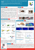
While MSI holds immense potential for advancing our understanding of spider metabolomics, several challenges needed to be addressed. In this context, the sample preparation of spider slices constitutes one of the main difficulties, the spider being made of very soft tissues and large proportion of biological fluids. Obtaining histological slices of Steatoda nobilis, without degrading the tissue or disturbing the internal anatomy represented a real challenge, that has been solved by the mean of a gelatine-based sample fixation. Organs specific metabolites such as lipids and amino acids allowed to rapidly identify various organs of the spiders, such as the silk glands, the ovaries, the venom glands or again the nervous systems. The second issue was linked to the quantity of data generated in each image. Each MS analysis yields to several thousands of peaks, each capable of providing a molecular image. To reduce the data treatment, ions were classified into structural families using Kendrick mass defect plots. Kendrick mass defect (KMD) analysis is widely used for helping the detection and identification of chemically related compounds based on exact mass measurements. Using this approach, we were able to link ions to molecules and localize these molecules into the spider body increasing the knowledge about the spider anatomy, at the molecular level.