Abstract :
[en] Introduction
The locus coeruleus (LC) is the main source of norepinephrine (NE) in the brain and sends monosynaptic projections to most of the brain (Szabadi, 2013). It contributes to multiple processes such as cognition and arousal (Mather & Harley, 2016). The LC further contributes heavily to the transition between sleep and wakefulness, and between slow wave sleep (SWS) and rapid eye movement sleep (REMS) (Cirelli, Tononi 2005). Through the local release of NE, it governs the synchrony of brain activity and is tightly linked to the oscillatory modes found during sleep, such as sleep spindles and slow waves in animals (Eschenko et al., 2012). While the LC appears to be an important structure for sleep, only limited imaging studies evaluated whether the LC is related to sleep variability due to the difficulty of imaging such a small size nucleus in vivo (Keren et al., 2009).
We aimed to investigate the link between LC activity during wakefulness and electroencephalogram (EEG) features of sleep using ultra-high-field 7-Tesla MRI. To trigger a response of the LC, we used a perceptual rivalry task. Subjects saw an ambiguous stimulus which could trigger spontaneous switches between two perceptions of the same image (Blake & Logothetis, 2002). Switches are considered to recruit bottom-up and top-down attentional processes associated with activity of the LC (Einhauser et al., 2008; Murphy et al., 2014).
Methods
50 healthy volunteers, including 33 young (age: 22.27y ± 3.21y; 29 women) and 17 late middle-aged (age: 61.35y ± 5.19y; 12 women) individuals completed the protocol. They first underwent a structural 7T MRI session, which allowed collecting high-resolution whole-brain T1-weighted images as well as a sequence dedicated to acquire 6cm LC slab. The latter was used to create in the brain space of each participant an individual LC-mask. All individual LC-masks were gathered into a probabilistic LC-mask in a standardized group brain space.
Following 1 week of regular sleep times, participants completed an fMRI session in the morning, 2h to 3h after wake-up time, during which they were administered the perceptual rivalry task (TR= 2.34s; voxel size 1.4x1.4x1.4 mm³). Participant’s habitual and baseline sleep was recorded in-lab under EEG to extract 4 sleep features of interest depicting some of the most canonical characteristics of sleep (i.e., cumulated power of the theta frequency band during REMS, slow wave energy (SWE), sleep onset latency, and REMS percentage).
We first conducted a general linear model with the Statistical Parametric Mapping 12 package (SPM12) over the entire brain. We then extracted the activity estimates over individual LC masks and then conducted generalized linear mixed models (GLMMs) to test for associations between the activity of the LC and EEG features of sleep, including age, sex, BMI, and total sleep time as covariates.
Results
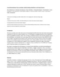
Perceptual switches were associated with increased activation in the left LC after controlling for age, sex, BMI, and total sleep time (whole brain FDR-corrected p<0.01; t > 3.055) (Fig1). This supported the extraction of individual LC activity in the individual space to associate with sleep EEG metrics. GLMMs on the LC activity and sleep EEG metrics revealed a positive association between the bilateral LC activity and SWE (cumulated EEG power over the .5-4Hz frequency band) (p = 0.02, F= 5.1), which reflects SWS intensity. A similar positive association was found between the LC activity and EEG cumulated power of the theta frequency band (4-8Hz) during REMS (p = 0.03, F=4.8), which is the dominant oscillatory mode of REMS. No other significant associations were detected.
Conclusions
These results show that a greater LC activity during wakefulness is associated with a more intense SWS and REMS. Future analyses will consider the relationship between LC activity and sleep microstructure.
Funding: FNRS Belgium, ULiège, FEDER, Alzheimer Foundation (SAO-FRA), Wallonia-Brussels federation
References
Blake, R., & Logothetis, N. K. (2002). Visual competition. Nature Reviews Neuroscience, 3(1), 13–21.
Einhauser, W., Stout, J., Koch, C., & Carter, O. (2008). Pupil dilation reflects perceptual selection and predicts subsequent stability in perceptual rivalry. Proceedings of the National Academy of Sciences, 105(5), 1704–1709. https://doi.org/10.1073/pnas.0707727105
Eschenko, O., Magri, C., Panzeri, S., & Sara, S. J. (2012). Noradrenergic neurons of the locus coeruleus are phase locked to cortical up-down states during sleep. Cerebral Cortex, 22(2), 426–435. https://doi.org/10.1093/cercor/bhr121
Keren, N. I., Lozar, C. T., Harris, K. C., Morgan, P. S., & Eckert, M. A. (2009). In vivo mapping of the human locus coeruleus. NeuroImage, 47(4), 1261–1267. https://doi.org/https://doi.org/10.1016/j.neuroimage.2009.06.012
Mather, M., & Harley, C. W. (2016). The Locus Coeruleus: Essential for Maintaining Cognitive Function and the Aging Brain. Trends in Cognitive Sciences, 20(3), 214–226. https://doi.org/10.1016/j.tics.2016.01.001
Murphy, P. R., O’Connell, R. G., O’Sullivan, M., Robertson, I. H., & Balsters, J. H. (2014). Pupil diameter covaries with BOLD activity in human locus coeruleus. Human Brain Mapping, 35(8), 4140–4154. https://doi.org/10.1002/hbm.22466
Scammell, T. E., Arrigoni, E., & Lipton, J. O. (2017). Neural Circuitry of Wakefulness and Sleep. Neuron, 93(4), 747–765. https://doi.org/10.1016/j.neuron.2017.01.014
Szabadi, E. (2013). Functional neuroanatomy of the central noradrenergic system. Journal of Psychopharmacology, 27(8), 659–693. https://doi.org/10.1177/0269881113490326