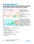
[en] The process of implantation and the cellular interactions at the embryo-maternal interface are intrinsically difficult to analyze, as the implanting embryo is concealed by the uterine tissues. Therefore, the mechanisms mediating the interconnection of the embryo and the mother are poorly understood. Here, we established a 3D biomimetic culture environment that harbors the key features of the murine implantation niche. This culture system enabled direct analysis of trophoblast invasion and revealed the first embryonic interactions with the maternal vasculature. We found that implantation is mediated by the collective migration of penetrating strands of trophoblast giant cells, which acquire the expression of vascular receptors, ligands, and adhesion molecules, assembling a network for communication with the maternal blood vessels. In particular, Pdgf signaling cues promote the establishment of the heterologous contacts. Together, the biomimetic platform and our findings thereof elucidate the hidden dynamics of the early interactions at the implantation site.
Disciplines :
Biochemistry, biophysics & molecular biology
Author, co-author :
Govindasamy, Niraimathi; Embryonic Self-Organization research group, Max Planck Institute for Molecular Biomedicine, Röntgenstraße 20, 48149 Münster, Germany
Long, Hongyan; Bioactive Materials Laboratory, Max Planck Institute for Molecular Biomedicine, Röntgenstraße 20, 48149 Münster, Germany
Jeong, Hyun-Woo; Department of Tissue Morphogenesis, Max Planck Institute for Molecular Biomedicine, Röntgenstraße 20, 48149 Münster, Germany
Raman, Ratish ; Université de Liège - ULiège > GIGA > GIGA I3 - Laboratory for Organogenesis and Regeneration ; Embryonic Self-Organization research group, Max Planck Institute for Molecular Biomedicine, Röntgenstraße 20, 48149 Münster, Germany
Özcifci, Burak; Center for Nanotechnology (CeNTech) und Physikalisches Institut Westfälische Wilhelms-Universität Münster Wilhelm-Klemm-Strasse 10, 48149 Münster, Germany
Probst, Simone; Institute of Experimental and Clinical Pharmacology and Toxicology, Faculty of Medicine, and Signaling Research Centers BIOSS and CIBSS, University of Freiburg, Germany
Arnold, Sebastian J; Institute of Experimental and Clinical Pharmacology and Toxicology, Faculty of Medicine, and Signaling Research Centers BIOSS and CIBSS, University of Freiburg, Germany
Riehemann, Kristina; Center for Nanotechnology (CeNTech) und Physikalisches Institut Westfälische Wilhelms-Universität Münster Wilhelm-Klemm-Strasse 10, 48149 Münster, Germany
Ranga, Adrian; Laboratory of Bioengineering and Morphogenesis, Department of Mechanical Engineering, KU Leuven, Leuven, Belgium
Adams, Ralf H; Department of Tissue Morphogenesis, Max Planck Institute for Molecular Biomedicine, Röntgenstraße 20, 48149 Münster, Germany
Trappmann, Britta; Bioactive Materials Laboratory, Max Planck Institute for Molecular Biomedicine, Röntgenstraße 20, 48149 Münster, Germany. Electronic address: britta.trappmann@mpi-muenster.mpg.de
Bedzhov, Ivan; Embryonic Self-Organization research group, Max Planck Institute for Molecular Biomedicine, Röntgenstraße 20, 48149 Münster, Germany. Electronic address: ivan.bedzhov@mpi-muenster.mpg.de
Language :
English
Title :
3D biomimetic platform reveals the first interactions of the embryo and the maternal blood vessels.
We thank Prof. Dr. Hans R. Schöler, Prof. Dr. Dietmar Vestweber, Prof. Dr. Friedemann Kiefer, and Dr. Mara Pitulescu for constructive discussions and suggestions and for providing access to key infrastructure, equipment and reagents; Dr. Rodrigo Dieguez-Hurtado for the protocols and technical advice for sorting endothelial cells; Prof. Dr. Christopher S. Chen for providing the molds for the device housing; Dr. Martin Stehling (Flow cytometry unit, MPI-MB), Dr. Stefan Volkery and Malte Stasch (BioOptic service unit, MPI-MB), Susanne Martin, and Anna Plogmann (Animal facility, MPI-MB), Heike Brinkmann, Saskia Winter, and Verena Stegemann for the excellent technical support; Fei Chen and all members of Bedzhov and Trappmann labs for the constructive discussions and suggestions, and Dr. Celeste Brennecka for proofreading the manuscript. Funding: This work was supported by the German Research Foundation (DFG) Emmy Noether grant ( BE 5800/1-1 ) and the Collaborative Research Center 1348 ‘Dynamic Cellular Interfaces’ grant ( 1348/1, B09 ) to I.B.; the Cells-In-Motion Cluster of Excellence (CiM) Flexible Funds grant ( FF-2017-04 ) to I.B. and B.T.; Collaborative Research Center 1348 ‘Dynamic Cellular Interfaces’ grant ( 1348/1 , A07) to B.T.; ( AR 732/3-1 , AR 732/2-1 , SFB850 (A03) ), Germany's Excellence Strategy —EXC-2189—project ID: 390939984 to S.J.A., the Leducq Foundation grant to H-W.J. and R.H.A. and CiM Pilot project grant ( PP-2020-10 ) to N.G. N.G. is supported by the International Max Planck Research School —Molecular Biology and Medicine, Münster, Germany. H.L. is a member of the integrated research training group of the Collaborative Research Center 1348
Alexander, S., Koehl, G.E., Hirschberg, M., Geissler, E.K., Friedl, P., Dynamic imaging of cancer growth and invasion: a modified skin-fold chamber model. Histochem. Cell Biol. 130 (2008), 1147–1154.
Anders, S., Pyl, P.T., Huber, W., HTSeq–a Python framework to work with high-throughput sequencing data. Bioinformatics 31 (2015), 166–169.
Arnold, S.J., Hofmann, U.K., Bikoff, E.K., Robertson, E.J., Pivotal roles for eomesodermin during axis formation, epithelium-to-mesenchyme transition and endoderm specification in the mouse. Development 135 (2008), 501–511.
Artus, J., Panthier, J.J., Hadjantonakis, A.-K., A role for PDGF signaling in expansion of the extra-embryonic endoderm lineage of the mouse blastocyst. Development 137 (2010), 3361–3372.
Ault, P., Kantarjian, H., O'Brien, S., Faderl, S., Beran, M., Rios, M.B., Koller, C., Giles, F., Keating, M., Talpaz, M., Cortes, J., Pregnancy among patients with chronic myeloid leukemia treated with imatinib. J. Clin. Oncol. 24 (2006), 1204–1208.
Babiarz, B., Romagnano, L., Afonso, S., Kurila, G., Localization and expression of fibronectin during mouse decidualization in vitro: mechanisms of cell:matrix interactions. Dev. Dyn. 206 (1996), 330–342.
Bedzhov, I., Alotaibi, H., Basilicata, M.F., Ahlborn, K., Liszewska, E., Brabletz, T., Stemmler, M.P., Adhesion, but not a specific cadherin code, is indispensable for ES cell and induced pluripotency. Stem Cell Res. 11 (2013), 1250–1263.
Bedzhov, I., Leung, C.Y., Bialecka, M., Zernicka-Goetz, M., In vitro culture of mouse blastocysts beyond the implantation stages. Nat. Protoc. 9 (2014), 2732–2739.
Bedzhov, I., Zernicka-Goetz, M., Self-organizing properties of mouse pluripotent cells initiate morphogenesis upon implantation. Cell 156 (2014), 1032–1044.
Blankenship, T.N., Enders, A.C., Expression of platelet-endothelial cell adhesion molecule-1 (PECAM) by macaque trophoblast cells during invasion of the spiral arteries. Anat. Rec. 247 (1997), 413–419.
Boomsma, C.M., Kavelaars, A., Eijkemans, M.J., Lentjes, E.G., Fauser, B.C., Heijnen, C.J., Macklon, N.S., Endometrial secretion analysis identifies a cytokine profile predictive of pregnancy in IVF. Hum. Reprod. 24 (2009), 1427–1435.
Cross, J.C., Werb, Z., Fisher, S.J., Implantation and the placenta: key pieces of the development puzzle. Science 266 (1994), 1508–1518.
Damsky, C.H., Fisher, S.J., Trophoblast pseudo-vasculogenesis: faking it with endothelial adhesion receptors. Curr. Opin. Cell Biol. 10 (1998), 660–666.
Damsky, C.H., Fitzgerald, M.L., Fisher, S.J., Distribution patterns of extracellular matrix components and adhesion receptors are intricately modulated during first trimester cytotrophoblast differentiation along the invasive pathway, in vivo. J. Clin. Invest. 89 (1992), 210–222.
Ehrbar, M., Rizzi, S.C., Hlushchuk, R., Djonov, V., Zisch, A.H., Hubbell, J.A., Weber, F.E., Lutolf, M.P., Enzymatic formation of modular cell-instructive fibrin analogs for tissue engineering. Biomaterials 28 (2007), 3856–3866.
Fan, R., Kim, Y.S., Wu, J., Chen, R., Zeuschner, D., Mildner, K., Adachi, K., Wu, G., Galatidou, S., Li, J., et al. Wnt/beta-catenin/Esrrb signalling controls the tissue-scale reorganization and maintenance of the pluripotent lineage during murine embryonic diapause. Nat. Commun., 11, 2020, 5499.
Friedl, P., Gilmour, D., Collective cell migration in morphogenesis, regeneration and cancer. Nat. Rev. Mol. Cell Biol. 10 (2009), 445–457.
Govindasamy, N., Duethorn, B., Oezgueldez, H.O., Kim, Y.S., Bedzhov, I., Test-tube embryos - mouse and human development in vitro to blastocyst stage and beyond. Int. J. Dev. Biol. 63 (2019), 203–215.
Haimovici, F., Anderson, D.J., Effects of growth factors and growth factor-extracellular matrix interactions on mouse trophoblast outgrowth in vitro. Biol. Reprod. 49 (1993), 124–130.
Hasford, J., Pfirrmann, M., Hochhaus, A., How long will chronic myeloid leukemia patients treated with imatinib mesylate live?. Leukemia 19 (2005), 497–499.
Kim, D., Pertea, G., Trapnell, C., Pimentel, H., Kelley, R., Salzberg, S.L., TopHat2: accurate alignment of transcriptomes in the presence of insertions, deletions and gene fusions. Genome Biol., 14, 2013, R36.
Klauber, N., Rohan, R.M., Flynn, E., D'Amato, R.J., Critical components of the female reproductive pathway are suppressed by the angiogenesis inhibitor AGM-1470. Nat. Med. 3 (1997), 443–446.
Lindahl, P., Johansson, B.R., Levéen, P., Betsholtz, C., Pericyte loss and microaneurysm formation in PDGF-B-deficient mice. Science 277 (1997), 242–245.
Love, M.I., Huber, W., Anders, S., Moderated estimation of fold change and dispersion for RNA-seq data with DESeq2. Genome Biol., 15, 2014, 550.
Ma, W.-G., Song, H., Das, S.K., Paria, B.C., Dey, S.K., Estrogen is a critical determinant that specifies the duration of the window of uterine receptivity for implantation. Proc. Natl. Acad. Sci. USA 100 (2003), 2963–2968.
Muzumdar, M.D., Tasic, B., Miyamichi, K., Li, L., Luo, L., A global double-fluorescent Cre reporter mouse. Genesis 45 (2007), 593–605.
Nakayama, H., Scott, I.C., Cross, J.C., The transition to endoreduplication in trophoblast giant cells is regulated by the mSNA zinc finger transcription factor. Dev. Biol. 199 (1998), 150–163.
Nancy, P., Tagliani, E., Tay, C.-S., Asp, P., Levy, D.E., Erlebacher, A., Chemokine gene silencing in decidual stromal cells limits T cell access to the maternal-fetal interface. Science 336 (2012), 1317–1321.
Nguyen, D.-H.T., Stapleton, S.C., Yang, M.T., Cha, S.S., Choi, C.K., Galie, P.A., Chen, C.S., Biomimetic model to reconstitute angiogenic sprouting morphogenesis in vitro. Proc. Natl. Acad. Sci. USA 110 (2013), 6712–6717.
Novartis. Investigators Brochure. STI571 (formerly CGP57148B) and data on file, Novartis Pharma AG, Basel, Switzerland.
Pandya, P., Orgaz, J.L., Sanz-Moreno, V., Modes of invasion during tumour dissemination. Mol. Oncol. 11 (2017), 5–27.
Probst, S., Daza, R.A., Bader, N., Hummel, J.F., Weiß, M., Tanriver, Y., Hevner, R.F., Arnold, S.J., A dual-fluorescence reporter in the Eomes locus for live imaging and medium-term lineage tracing. Genesis, 55, 2017, e23043.
Pye, S.M., Cortes, J., Ault, P., Hatfield, A., Kantarjian, H., Pilot, R., Rosti, G., Apperley, J.F., The effects of imatinib on pregnancy outcome. Blood 111 (2008), 5505–5508.
Rai, A., Cross, J.C., Development of the hemochorial maternal vascular spaces in the placenta through endothelial and vasculogenic mimicry. Dev. Biol. 387 (2014), 131–141.
Ramilowski, J.A., Goldberg, T., Harshbarger, J., Kloppmann, E., Lizio, M., Satagopam, V.P., Itoh, M., Kawaji, H., Carninci, P., Rost, B., Forrest, A.R.R., A draft network of ligand-receptor-mediated multicellular signalling in human. Nat. Commun., 6, 2015, 7866.
Ranga, A., Girgin, M., Meinhardt, A., Eberle, D., Caiazzo, M., Tanaka, E.M., Lutolf, M.P., Neural tube morphogenesis in synthetic 3D microenvironments. Proc. Natl. Acad. Sci. USA 113 (2016), E6831–E6839.
Reus, A.D., El-Harbachi, H., Rousian, M., Willemsen, S.P., Steegers-Theunissen, R.P.M., Steegers, E.A.P., Exalto, N., Early first-trimester trophoblast volume in pregnancies that result in live birth or miscarriage. Ultrasound Obstet. Gynecol. 42 (2013), 577–584.
Rider, V., Carlone, D.L., Witrock, D., Cai, C., Oliver, N., Uterine fibronectin mRNA content and localization are modulated during implantation. Dev. Dyn. 195 (1992), 1–14.
Riedl, J., Crevenna, A.H., Kessenbrock, K., Yu, J.H., Neukirchen, D., Bista, M., Bradke, F., Jenne, D., Holak, T.A., Werb, Z., et al. Lifeact: a versatile marker to visualize F-actin. Nat. Methods 5 (2008), 605–607.
Russ, A.P., Wattler, S., Colledge, W.H., Aparicio, S.A.J.R., Carlton, M.B.L., Pearce, J.J., Barton, S.C., Surani, M.A., Ryan, K., Nehls, M.C., et al. Eomesodermin is required for mouse trophoblast development and mesoderm formation. Nature 404 (2000), 95–99.
Russell, M.A., Carpenter, M.W., Akhtar, M.S., Lagattuta, T.F., Egorin, M.J., Imatinib mesylate and metabolite concentrations in maternal blood, umbilical cord blood, placenta and breast milk. J. Perinatol. 27 (2007), 241–243.
Salem, W., Li, K., Krapp, C., Ingles, S.A., Bartolomei, M.S., Chung, K., Paulson, R.J., Nowak, R.A., McGinnis, L.K., Imatinib treatments have long-term impact on placentation and embryo survival. Sci. Rep., 9, 2019, 2535.
Simmons, D.G., Fortier, A.L., Cross, J.C., Diverse subtypes and developmental origins of trophoblast giant cells in the mouse placenta. Dev. Biol. 304 (2007), 567–578.
Sutherland, A.E., Calarco, P.G., Damsky, C.H., Developmental regulation of integrin expression at the time of implantation in the mouse embryo. Development 119 (1993), 1175–1186.
Thomas, T., Dziadek, M., Expression of laminin and nidogen genes during the postimplantation development of the mouse placenta. Biol. Reprod. 49 (1993), 1251–1259.
Torry, D.S., Leavenworth, J., Chang, M., Maheshwari, V., Groesch, K., Ball, E.R., Torry, R.J., Angiogenesis in implantation. J. Assist. Reprod. Genet. 24 (2007), 303–315.
Vićovac, L., Jones, C.J., Aplin, J.D., Trophoblast differentiation during formation of anchoring villi in a model of the early human placenta in vitro. Placenta 16 (1995), 41–56.
Wang, J., Armant, D.R., Integrin-mediated adhesion and signaling during blastocyst implantation. Cells Tissues Organs 172 (2002), 190–201.
Wang, Y., Wang, R., Zhang, S., Song, S., Jiang, C., Han, G., Wang, M., Ajani, J., Futreal, A., Wang, L., iTALK: an R package to characterize and illustrate intercellular communication. bioRxiv, 2019, 10.1101/507871.
Winderlich, M., Keller, L., Cagna, G., Broermann, A., Kamenyeva, O., Kiefer, F., Deutsch, U., Nottebaum, A.F., Vestweber, D., VE-PTP controls blood vessel development by balancing Tie-2 activity. J. Cell Biol. 185 (2009), 657–671.
Zhou, J.X., Taramelli, R., Pedrini, E., Knijnenburg, T., Huang, S., Extracting intercellular signaling network of cancer tissues using ligand-receptor expression patterns from whole-tumor and single-cell transcriptomes. Sci. Rep., 7, 2017, 8815.
Zhou, Y., Fisher, S.J., Janatpour, M., Genbacev, O., Dejana, E., Wheelock, M., Damsky, C.H., Human cytotrophoblasts adopt a vascular phenotype as they differentiate. A strategy for successful endovascular invasion?. J. Clin. Invest. 99 (1997), 2139–2151.
Zhu, J.-Y., Pang, Z.-J., Yu, Y.-H., Regulation of trophoblast invasion: the role of matrix metalloproteinases. Rev. Obstet. Gynecol. 5 (2012), e137–e143.