Education and training; Computer Applications-Detection; diagnosis; Cone beam CT; Head and neck
Abstract :
[en] Purpose or Learning Objective
To propose different diagnostical steps regarding the majority and the most frequent dentomaxillary lesions. The first step is lesional description, the second precise lesional topography, the third discuss a sex predominance and age of diagnosis, and the fourth is a discussion about specificity of semiological signs.
Methods or Background
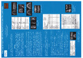
For many years the gold standard in dentomaxillary imaging were the intraoral and panoramic radiographies, in some cases the CT. The Cone Beam Computed Tomography (CBCT) put maxillofacial imaging on higher level than before. The small field of intraoral radiography and two-dimensional panoramic radiography don’t allow a correct check-up of many neoplastic, systemic, inflammatory and sometimes of large infectious pathologies. The CBCT with its possibilities of three-dimensional and multiplanar reconstructions, with its higher resolution than a CT, allows more accurate differential diagnosis of dentomaxillary pathologies. In certains conditions such as the malignant pathologies we can need a MRI and histological analysis.
In some countries there are radiologists making dentomaxillary imaging and in others dentists. Because of various pathologies specifically linked to maxillary bones use of dentomaxillary CBCT is found to be more complex than a panoramic or an oral radiography for dentists and not at all easier for radiologists. In both cases it is the question of specific training for these pathologies suspected by CBCT.
Results or Findings
Diagnosis is always oriented by the clinic aided by CBCT, more rarely by CT or MRI and finally often completed by histological analysis especially in tumoral pathologies.
Disciplines :
Radiology, nuclear medicine & imaging
Author, co-author :
MILICEVIC, Mladen ; Centre Hospitalier Universitaire de Liège - CHU > Département de Physique Médicale > Service médical de radiodiagnostic
Language :
English
Title :
Four steps for rapid differential diagnosis of dentomaxillary pathologies
Alternative titles :
[en] Quatre pas pour un diagnostic différentiel rapide des pathologies dento-maxillaires