Abstract :
[en] Purpose or Objective
Neoadjuvant radiotherapy (NeoRT) improves tumor local control and facilitates tumor resection in many cancers. We hypothesized anti-cancer treatments (i.e. radiotherapy) modify tumor microenvironment and could potentially impact distant metastases occurrence. Previously, we developed a pre-clinical model demonstrating an impact of NeoRT schedule and the timing of surgery on metastatic spreading (Leroi et al. Oncotarget 2015). Here, we aim to identify by fMRI noninvasive markers reflecting NeoRT related tumor microenvironment modifications that could predict the best timing for performing surgery and avoiding tumor spreading.
Material and Methods
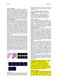
To briefly delineate the NeoRT model, MDA-MB 231 tumor cells implanted in the flank of SCID mice were locally irradiated with 2x5Gy when tumor reached 100mm3 and then surgically removed at different time points. We performed fMRI, Diffusion Weighted (DW) and Dynamic Contract enhancement (DCE) – MRI, before RT and every 2 days between RT and surgery. We acquired 8 slices of 1 mm thickness and 0.5 mm gap with an “in plane voxel resolution” of 0.5 mm. For DW-MRI, we performed FSEMS (Fast Spin Echo MultiSlice) sequences, with 9 different Bvalue (from 40 to 1000) and B0. We performed IVIM (IntraVoxel Incoherent Motion) analysis to obtain information on intravascular diffusion, related to perfusion (F: perfusion factor) and subsequently tumor vessels perfusion. For DCE-MRI, we performed a T1 mapping with multiple TR and DCE acquisition with 200 repetitions of 3 sec each and gadolinium IV injection after 10 repetitions. We performed semi-quantitative analysis. We validated tumor perfusion by immunochemistry with injection of FITC-dextran IV 3 min before surgery and CD31 labelling. Human Ki67 was used for lung metastases labelling and quantification.
Results
After the tumor irradiation, we observed a significant and transient increase at day 6 (60% of the basal value (n=6, p<0,05)) of F and D* parameters related to perfusion. The other parameters of the DW-MRI, ADC and D presented no modifications. The sham irradiated tumors used as control showed no modifications of all fMRI parameters. At the same timing, 6 days post-radiotherapy, DCE-MRI significantly demonstrated a WhashinSlope (n=13, p<0,05) increase. Immunochemistry confirmed the increase of tumor perfusion when surgery is performed at day 6. The sham irradiated tumors never demonstrated such changes. Finally, when surgery is performed on tumor increased perfusion measured by fMRI, it demonstrated a burst of lung metastasis compared to the other timings.
Conclusion
We showed a significant difference in perfusion-related parameters with fMRI and immunochemistry at a specific time point after NeoRT. These modifications are correlated with an increase of metastasis spreading related to surgery procedure. These results open new perspectives in the personalized medicine and MRI guided surgery timing after NeoRT.