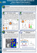
[en] During the last decade, spectroscopic cellular imaging has increasingly become a powerful characterization method for the study of all kind of cellular-related disease. In this context, Surface-Enhanced Raman Spectroscopy (SERS) is a powerful technique with many advantages over other spectroscopic techniques such as fluorescence or infrared spectroscopy. The SERS effect is capable of providing huge enhancement factors, highly increasing the sensitivity of Raman spectroscopy. One interesting approach is to use so called “SERS nanoprobes” to selectively target and detect a particular receptor, protein, DNA sequence, etc. In the area of SERS detection of cancerous cells, the target of these nanoprobes is usually a surface receptor that is characteristic of the cancerous state of the cell, or that is overexpressed in a cancerous cell in comparison to a healthy situation. In this study, we investigated the targeting of the Folate Receptor alpha (FRα) which is overexpressed in several cancers (ovarian and lung adenocarcinoma, among others) and can therefore be used for the imaging and detection of cancerous cells. We focused on the development of highly sensitive SERS nanoprobes, combining bimetallic nanoparticles and resonant Raman-active molecules.
We successfully imaged Folate Receptor α at the surface of two kinds of cancerous cells (KB and PC-3) thanks to the high confocality of the Raman micro-spectrometer. Moreover, we were able to distinguish these two kinds of cells by measuring the SERS intensity coming from each cell population, since both kinds of cells have a different expression level of the FRα receptor and will therefore accumulate different amounts of nanoprobes. Our approach provides new perspectives toward the discrimination of cancerous and healthy cells in real samples.
Research Center/Unit :
Laboratory of Inorganic Analytical Chemistry Laboratory of Culture of Mammalian Cells
Disciplines :
Chemistry
Author, co-author :
Verdin, Alexandre ; Université de Liège - ULiège > Département de chimie (sciences) > Département de chimie (sciences)
Malherbe, Cédric ; Université de Liège - ULiège > Département de chimie (sciences) > Chimie analytique inorganique
Cambroisier, Florie ; Université de Liège - ULiège > Département des sciences biomédicales et précliniques > Histologie - Cytologie
Bertrand, Virginie ; Université de Liège - ULiège > Département de chimie (sciences) > Département de chimie (sciences)
De Pauw-Gillet, Marie-Claire ; Université de Liège - ULiège > Département des sciences biomédicales et précliniques > Histologie - Cytologie
Eppe, Gauthier ; Université de Liège - ULiège > Département de chimie (sciences) > Chimie analytique inorganique
Language :
English
Title :
Development of highly sensitive SERS nanoprobes for cellular imaging of Folate Receptor alpha