[en] Introduction
In human medicine, far lateral lumbar disc extrusion (FLLDE) represents 7 to 12% of all disc herniations and MRI is the method of choice for diagnosis. MRI findings of a FLLDE has been reported in one dog and the aim of this case report is to describe computed tomographic (CT) findings of a FLLDE in a dog.
Methods
A six-year-old neutered female Beagle with a good general health status was presented with a month history of left hind limb pain with shivering of this limb. Clinical examination revealed a left hind proprioceptive deficit but no pain was elicited at palpation.
Results
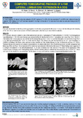
A CT scan pre- and post- contrast studies of the lumbosacral spine and a myelographic-CT were performed. A 1.3x0.6x0.3 cm homogeneous hyperattenuating (+/- 350 HU) ovoid structure was observed at the left lateral aspect of L6-L7 intervertebral disc space. The L6-L7 intervertebral disc nucleus pulposus was calcified but the annulus fibrosus had a normal non-calcified attenuation and seemed intact apart from a very thin hyperattenuating line next to the ovoid structure. This change was well circumscribed by an even hyperattenuating rim (1000HU) mimicking a thin dense cortex and was in close proximity with the annulus fibrosus and the left transverse process of L7. Perineural fat was not observed and contrast enhancement was visualized at the level of the left sixth lumbar nerve root with impingement and thickening of this root. No compression of the spinal cord was observed at this level on the myeloCT. These findings were suggestive of a dystrophic mineralization or an osteochondromatosis. FLLDE was considered less likely because of the almost normal appearance of the annulus fibrosus. At surgery some mixed gelified calcified material consistent with disc material was removed at the level of the left L6-L7 nerve root tract and disc fenestration was performed. The histological analysis confirmed the presence of degenerated herniated vertebral disk.
Discussion/Conclusions
In human medicine, MRI and CT scan are the main diagnostic modalities employed for diagnosis of FLLDE. In veterinary medicine, CT is often used to assess spinal diseases because of its availability. To the authors’ knowledge, CT findings of a FLLDE have not been described previously in dogs. Moreover, the thin dense cortex appearance surrounding the disc material was surprising. In conclusion, FLLDE should be included in the differential diagnosis of a calcified ovoid structure lateral to the spine even if the annulus fibrosus appears normal and if this structure is in close relation with the vertebral transverse process.
Research Center/Unit :
FARAH - Fundamental and Applied Research for Animals and Health - ULiège
Disciplines :
Veterinary medicine & animal health
Author, co-author :
Rizza, Maïlis ; Université de Liège > Dép. clinique des animaux de compagnie et des équidés (DCA) > Imagerie médicale
Bouvy, Bernard ; Université de Liège > Dép. clinique des animaux de compagnie et des équidés (DCA) > Chirurgie et clinique chirurgicale des petits animaux
Shimizu, Naomi ; Université de Liège > Dép. clinique des animaux de compagnie et des équidés (DCA) > Chirurgie et clinique chirurgicale des petits animaux
Heimann, Marianne; Laboratory Anapet SPRL Belgium
Bolen, Géraldine ; Université de Liège > Dép. clinique des animaux de compagnie et des équidés (DCA) > Imagerie médicale
Language :
English
Title :
Computed tomographic findings of a far lateral lumbar disc extrusion in a dog
Publication date :
01 September 2016
Event name :
European Veterinary Diagnostic Imaging annual scientific meeting 2016 (EVDI 2016)