[en] Introduction: The study of normal structures of the sheep’s brain is very important to understand pathological changes caused by the bluetongue virus in the fetus’s brain at various stages of the gestation. Bluetongue is an arthropod-borne viral disease of domestic and wild ruminants. The serotype 8 is responsible for outbreaks in Northern Europe in 2006. This virus causes lesions in the brain of fetuses as hydrancephaly and porencephaly. The aim of this work is to improve knowledge of anatomy and histology of the central nervous system of the sheep.
Methods: Seven heads of adult sheep and one from a fetus aged 4,5 months were used. All heads were first opened in the frontal area using bone’s saw and immerged in a formalin solution for 10 days. After a good fixation, the brains were extracted and sectioned. Transversal, frontal and sagittal sections were realized. The sections of two brains were stained with Berlin-blue and treated to be embedded in methylmetacrylate for gross morphology. The different parts of the 6 resting brains were then embedded in paraffin, cut and the histological sections were stained with haematoxylin/eosin, cresyl violet or by use of silver impregnation.
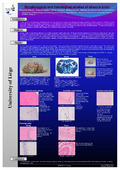
Results: Gross morphological examination of the brains embedded in methylmetacrylate showed the detailed anatomy of the different parts. The staining with haematoxylin/eosin permitted to differentiate the grey matter, the different nucleus and the layers of cerebral and cerebellum cortex. The cresyl violet technique permitted to visualize the Nissl bodies and the silver impregnation revealed nerve fibers. In the fetus brain, blood vessels were very numerous in the brainstem, the cerebellum and the cerebrum. The grey matter was less organized and looser.
Conclusion: This work establishes an anatomical and histological approach allowing future studies in ovine fetuses with and without brain lesions potentially caused by the bluetongue virus.